Outlook Best Doctors South 2025
Bagi Heart Centre — Expertise. Technology. Compassion.
Precision Coronary Imaging

Intravascular Imaging

Catheter-based imaging from inside a coronary artery that adds detailed information to angiography and can guide complex angioplasty and stent placement.

Overview

Understanding this service

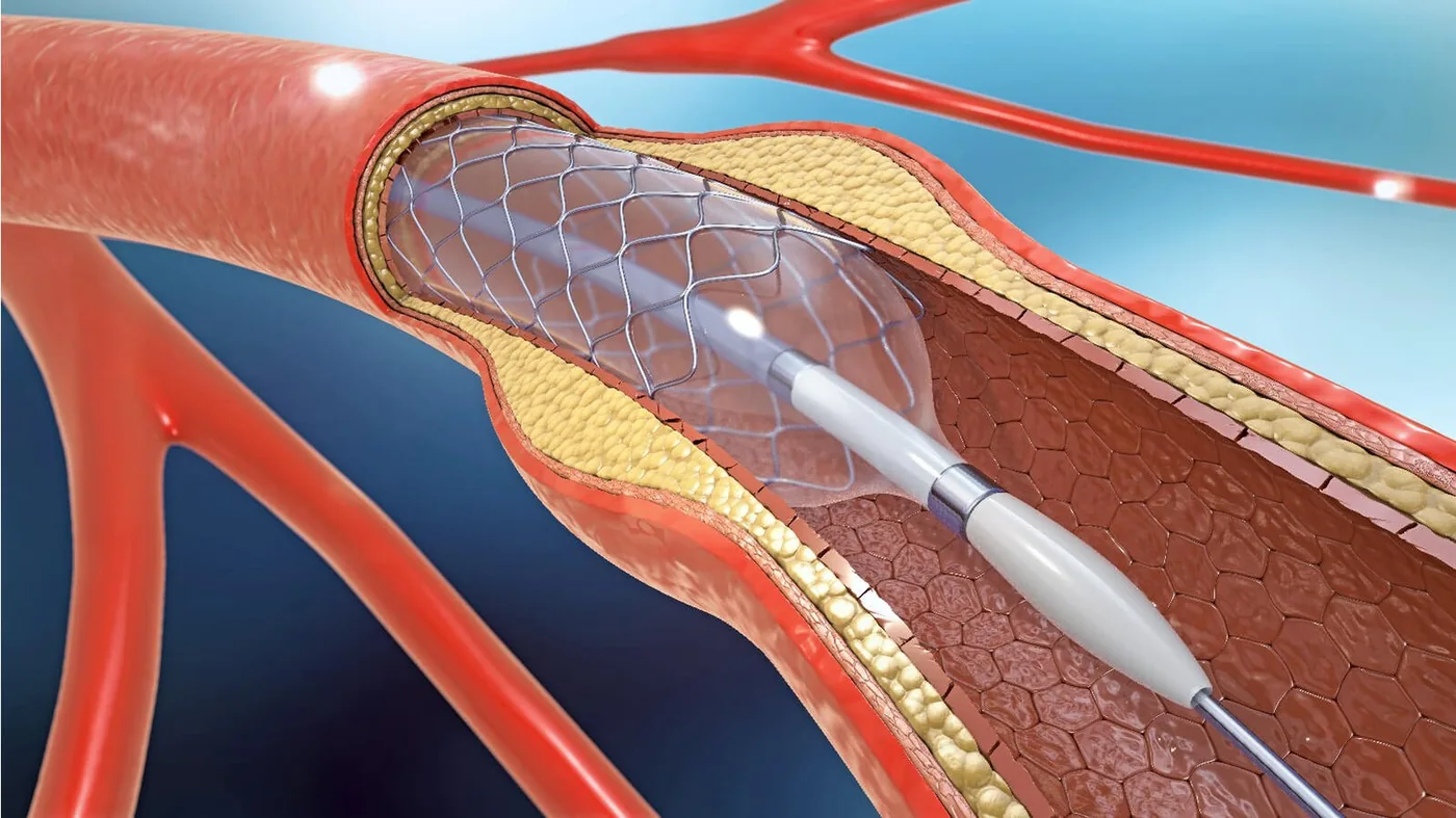
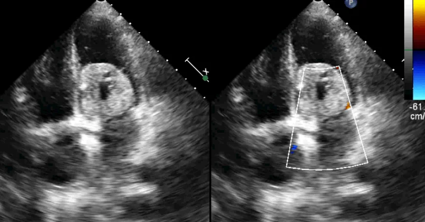
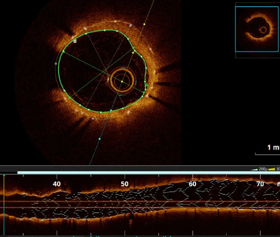
Intravascular imaging uses a very small imaging catheter inside a coronary artery to show the vessel wall and lumen in greater detail than angiography alone. Common techniques include intravascular ultrasound (IVUS) and optical coherence tomography (OCT).

It is usually performed during coronary angiography or angioplasty rather than as a separate operation. The images can help the cardiologist understand a lesion, measure the artery and assess how well a stent has expanded and contacted the vessel wall.

When is it recommended?

  • A coronary narrowing that is difficult to assess fully on angiography alone
  • Planning selected complex angioplasty procedures
  • Choosing an appropriate stent size and landing zone
  • Checking stent expansion, position and contact with the artery wall
  • Investigating selected cases of recurrent narrowing or a problem within a stent

Purpose and potential benefits

  • Provides a cross-sectional view from inside the coronary artery
  • Adds vessel measurements that can support treatment planning
  • Helps identify features that may not be clear on a two-dimensional angiogram
  • Allows the cardiologist to assess and optimise a newly placed stent
Intravascular Imaging — cardiac care at Dr. Bagi Heart Centre
Intravascular Imaging · Precision Coronary Imaging
What happens

How the pathway works.

Your exact pathway depends on your clinical assessment.

01

Angiographic access

The procedure uses the same wrist or groin arterial access established for coronary angiography or PCI, with local anaesthesia at the access site.

02

Imaging catheter placement

A guidewire is passed through the coronary artery and a small IVUS or OCT catheter is positioned across the area being assessed.

03

Image acquisition

The catheter records a controlled pullback through the artery. OCT generally requires contrast to clear blood briefly from the imaging field, while IVUS uses ultrasound.

04

Treatment guidance

The cardiologist reviews the images immediately and uses the findings to plan treatment or optimise a stent before removing the catheter.

Before & after

What patients can expect.

Intravascular imaging is performed in a catheterisation laboratory as part of an invasive coronary procedure.

Patients receive the same access-site preparation and monitoring used for angiography or angioplasty.

The imaging step adds detail to the procedure but does not replace the cardiologist's overall clinical assessment.

Aftercare follows the angiography or angioplasty plan, including access-site and medication instructions.

Why Dr. Bagi Heart Centre

One specialist view across diagnosis, treatment and follow-up.

Dr. Bagi combines interventional experience with non-invasive evaluation and hospital-based care coordination. Recommendations are made after reviewing symptoms, examination findings and relevant reports.

Clear consultationOptions explained in language you can use.
Procedural experience7,000+ interventional procedures recorded by the source site.
Connected evaluationTesting and follow-up aligned with your treatment pathway.
Continuity of careSupport before, during and after a major decision.
Questions, answered

Intravascular Imaging — frequently asked questions

General guidance only — your cardiologist will tailor advice to your health and reports.

An angiogram outlines the channel of the artery using X-ray and contrast. IVUS or OCT images the artery from within and can provide detailed cross-sectional information about the vessel and a stent.
Usually not. It is generally added during coronary angiography or angioplasty through the same arterial access.
No. The cardiologist uses it selectively when more detailed information could help plan or optimise treatment.
IVUS uses ultrasound from within the artery. OCT uses light and produces high-resolution images, usually with contrast used briefly during image capture. The choice depends on the clinical question.
Book a Consultation

Your heart deserves expert care — let's talk today

Book a consultation with Dr. Vithal D. Bagi on WhatsApp, or reserve a slot online. For emergencies, call our 24×7 line.